Shoulder Anatomy Diagram - Shoulder Anatomy Image Anatomy Drawing Diagram : Due to the inherent complexity of the shoulder joint, it is also particularly prone to problems.
Shoulder Anatomy Diagram - Shoulder Anatomy Image Anatomy Drawing Diagram : Due to the inherent complexity of the shoulder joint, it is also particularly prone to problems.. Plus, exercises for training them. Learn about these muscles, their origin and insertion points, and their functional anatomy. Ebraheim's educational animated video describes muscle anatomy of the shoulder girdle and anatomy of the shoulder joint.anatomy of the shoulder muscles a. A second joint in the shoulder is the junction of the collar bone with the shoulder blade, called the. The shoulder joint is formed where the humerus upper arm bone fits into the scapula shoulder blade like a ball and socket.
Starting with what is deepest, it goes: It is one of the most mobile joints in the human body, at the cost of joint stability. These muscles form the outer shape of the shoulder and underarm. The muscles of the shoulder bridge the transitions from the torso into the head/neck area and into the upper extremities of the arms and hands. Discuss tha agaonist/antagonist relationship of muscles.
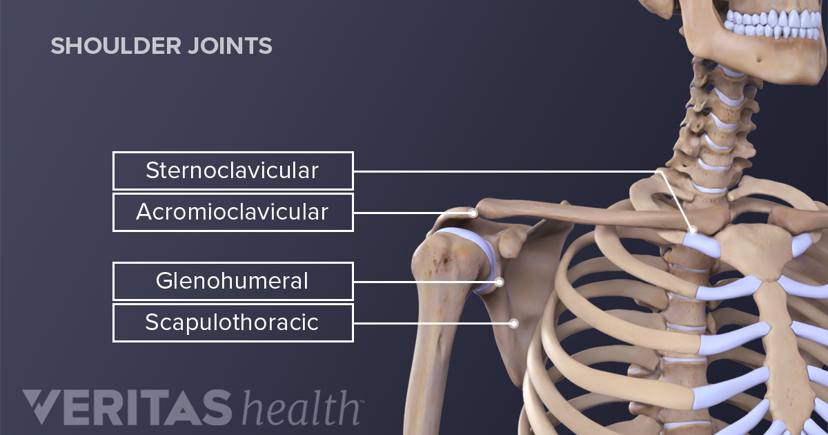
Shoulder Joint Human Anatomy Arm Face Hand Png Pngegg from e7.pngegg.com It is one of the most mobile joints in the human body, at the cost of joint stability. The glenohumeral joint is where the ball (humeral head) and the socket (the glenoid) meet. The shoulder is made up of two joints, the acromioclavicular joint and the glenohumeral joint. The shoulder joint is formed where the humerus (upper arm bone) fits into the scapula (shoulder blade), like a ball and socket. 13 461 shoulder anatomy stock photos pictures royalty free images istock. The shoulder has about eight muscles that attach to the scapula, humerus, and clavicle. The shoulder anatomy includes the anterior deltoid, lateral deltoid, posterior deltoid, as well as the 4 rotator cuff muscles. 17 photos of the diagram of shoulder muscles and tendons.
Human muscle system, the muscles of the human body that work the skeletal system, that are under voluntary control, and that are.
There are many nerves and blood vessels that supply the muscles and bones of the shoulder. Shoulder anatomy models help provide patients and students with a better understanding of how the shoulder joint functions as well as explaining common shoulder. These muscles form the outer shape of the shoulder and underarm. The anatomy of the shoulder. The shoulder is made up of two joints, the acromioclavicular joint and the glenohumeral joint. Ebraheim's educational animated video describes muscle anatomy of the shoulder girdle and anatomy of the shoulder joint.anatomy of the shoulder muscles a. Learn about these muscles, their origin and insertion points, and their functional anatomy. The following is an overview of the shoulder muscle anatomy. Shoulder anatomy images stock photos vectors. This diagram depicts shoulder anatomy muscles diagram.human anatomy diagrams show internal organs, cells, systems, conditions, symptoms and sickness information and/or tips for healthy living. Anatomy • free medical books. Find out in this anatomy of the shoulder quiz. Name this muscle the largest of the shoulder group.
Shoulder anatomy images stock photos vectors. 17 photos of the diagram of shoulder muscles and tendons. Bone, then ligaments of the joint capsule, with tendons and muscles on top. A second joint in the shoulder is the junction of the collar bone with the shoulder blade, called the. The most common shoulder injuries involve the muscles, ligaments, cartilage, and tendons, rather than the bones.
Anatomy Of The Human Shoulder Joint from www.verywellhealth.com Deltoides triangular refers to the front head of the. Groin muscles diagram groin muscles diagram elegant body areas. Bones in shoulder, ligaments of the shoulder joint, parts of the shoulder joint, shoulder anatomy, shoulder joints and muscles, shoulder structure anatomy, shoulder tendon anatomy, shoulder tendons ligaments, human muscles, bones in shoulder, ligaments of the shoulder joint, parts of. The most common shoulder injuries involve the muscles, ligaments, cartilage, and tendons, rather than the bones. Muscles of the shoulder can be subdivided into a variety of groups depending on origin, topography, function or innervation. Discuss tha agaonist/antagonist relationship of muscles. The following is an overview of the shoulder muscle anatomy. Name this muscle the largest of the shoulder group.
The acromioclavicular joint is where the acromion, part of the shoulder blade (scapula) and the collar bone (clavicle) meet.
17 photos of the diagram of shoulder muscles and tendons. However, more serious injuries, such as complete rotator cuff tears, may require surgical repair. The neck muscles, including the sternocleidomastoid and the trapezius, are responsible for the gross motor movement in the muscular system of the head and neck. The shoulder is one of the most sophisticated and complicated joints of the human body. Deltoides triangular refers to the front head of the. The most common shoulder injuries involve the muscles, ligaments, cartilage, and tendons, rather than the bones. The shoulder joint is formed where the humerus upper arm bone fits into the scapula shoulder blade like a ball and socket. The shoulder joint is the junction between the chest and the upper extremity. Starting with what is deepest, it goes: Shoulder anatomy models help provide patients and students with a better understanding of how the shoulder joint functions as well as explaining common shoulder. Muscles of the shoulder can be subdivided into a variety of groups depending on origin, topography, function or innervation. Ebraheim's educational animated video describes muscle anatomy of the shoulder girdle and anatomy of the shoulder joint.anatomy of the shoulder muscles a. Shoulder anatomy is formed by the union of three major bones including the humerus scapula and clavicle.
The following is an overview of the shoulder muscle anatomy. Rotator cuff injuries are very common, affecting over 3 million people in the united states every year. 13 461 shoulder anatomy stock photos pictures royalty free images istock. The muscles in the shoulder aid in a wide. Formerly called tendinitis, this is inflammation or irritation of a tendon that attaches to a bone.
Shoulder Joint Structure from embed.widencdn.net Discuss tha agaonist/antagonist relationship of muscles. Learn their origins/insertions, functions & exercises. The shoulder joint is the junction between the chest and the upper extremity. Male shoulder ligaments and biceps muscles isolated in skeleton labeled chart on white labeled human anatomy diagram of male shoulder ligaments, connective tissue and biceps muscles isolated within the skeletal system frontal anterior view on a white background. Muscles of the upper arm and shoulder blade human anatomy. Learn about these muscles, their origin and insertion points, and their functional anatomy. The muscles in the shoulder aid in a wide. The anatomy of the shoulder.
Numerous muscles help stabilize the three joints of.
This diagram depicts shoulder anatomy muscles diagram.human anatomy diagrams show internal organs, cells, systems, conditions, symptoms and sickness information and/or tips for healthy living. Most people with rotator cuff injuries can recover with rest and physical therapy. Bone, then ligaments of the joint capsule, with tendons and muscles on top. Discuss tha agaonist/antagonist relationship of muscles. Find out in this anatomy of the shoulder quiz. This diagram depicts diagram floating shoulder xray.human anatomy diagrams show internal organs, cells, systems, conditions, symptoms and sickness information and/or tips for healthy living. The most common shoulder injuries involve the muscles, ligaments, cartilage, and tendons, rather than the bones. Male shoulder ligaments and biceps muscles isolated in skeleton labeled chart on white labeled human anatomy diagram of male shoulder ligaments, connective tissue and biceps muscles isolated within the skeletal system frontal anterior view on a white background. The shoulder has about eight muscles that attach to the scapula, humerus, and clavicle. The anterior shoulder pain usually develops when injury or inflammation occurs in the tendons that are attached to the shoulder joint. The shoulder joint is the junction between the chest and the upper extremity. Scapula shoulder blade in shoulder pain workshop back 2 fitness. The following is an overview of the shoulder muscle anatomy.


/shoulder_pain_medreview-01-5c3b9f8546e0fb0001bdeaaa-d0a4923b7a3d441fb12d992c454a8ca7.png)


0 Komentar